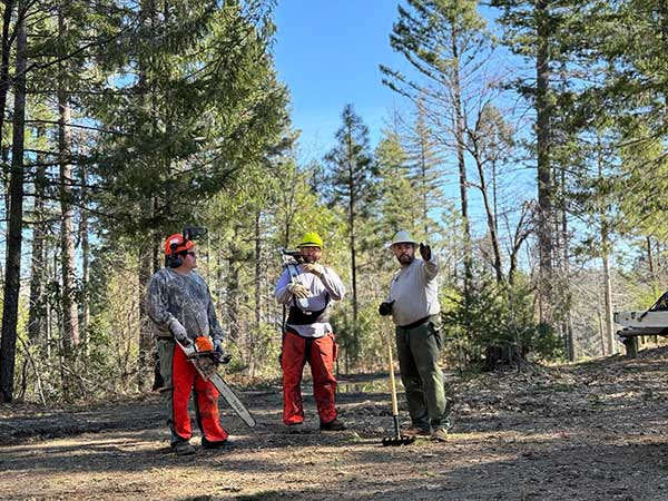
Upper Lake District recreation staff use chain saws and hand tools to clean up downed trees and debris in Deer Valley Campground. OHV trails and the campground will reopen Saturday, Mar. 16 after a storm damage closure lifts. USDA Forest Service photo by Andrew Avitt. MENDOCINO NATIONAL FOREST, Calif. — Mendocino National Forest staff are reopening off-highway vehicle, or OHV, trails and a campground after a closure lifts on Saturday, March 16, at midnight.
Upper Lake Ranger District OHV trails and the Deer Valley Campground had been closed due to extensive storm damage since mid-February.
Forest leadership mobilized volunteers and staff from recreation, fire, fuels and engineering to help with trail cleanup during the monthlong closure.
One campsite in the Deer Valley Campground will remain closed. A picnic table was destroyed by a fallen tree, and staff plan to restore the site when the ground is drier. Forest staff also continue to clear trails and conduct tread repair.
Forest officials caution visitors to be aware of their environment. Trees may continue to fall, and trail riders can expect to encounter downed trees on trails.
Roads in the forest can become impassable at any time due to downed trees, rockfall or slides. In higher elevations, roads remain impassable due to snow and ice.
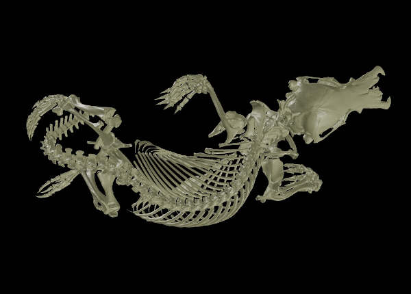
A 3D CT scan of a juvenile platypus from the collections of the UC Berkeley Museum of Vertebrate Zoology. (Image courtesy of MorphoSource and MVZ:Mamm: 32885) BERKELEY, Calif. — The Museum of Vertebrate Zoology, or MVZ, at the University of California, Berkeley, contains more than 300,000 vertebrate specimens — the majority of them reptiles and amphibians — preserved in alcohol and tucked away for current and future generations of scientists who want to study their anatomical and genetic diversity.
Now, those specimens are gradually gaining a new life online as part of an effort by 25 museums across the U.S. to obtain 3D scans of as many vertebrate groups as possible and make them available free to the general public in a searchable database.
A summary of the six-year project, called openVertebrate (oVert), was published this week in the journal BioScience, offering a glimpse of how the data might be used to ask new scientific questions and spur the development of innovative technology.
But scientists aren't the only ones who find the scans useful. Artists have used the 3D models to create realistic animal replicas, photographs of oVert specimens have been displayed as museum exhibits, and specimens have been incorporated into virtual reality headsets that give users the chance to interact with and manipulate them.
Carol Spencer, staff curator of herpetology in the MVZ, has a 3D-printed version of one specimen — the skull of a horned lizard — sitting on her desk. Anyone can access the 3D scans online at MorphoSource, download the data and send them to a 3D printer to produce their own skeletal models.
"You can actually print them and then use them in a classroom. We have lots of people using them for teaching in colleges or high schools," Spencer said.
Of the approximately 1,000 MVZ specimens scanned over the past six years through oVert, one — a juvenile Australian platypus, Ornithorhynchus anatinus — is the second most downloaded in the database.
"We've had this platypus in ethanol in a big tank, but it's never been loaned out. The only people who have ever gone to look at this are people that come here to our collection; it's maybe been looked at twice in its entire history here at MVZ. But in six years, it's been downloaded 320 times," Spencer said. "That's a huge expansion of use."
Spencer recently fielded a request from a professor at Towson University in Maryland to download CT scans for a course in which students compare the cranial anatomy of vertebrates and print 3D models for study.
"All of these specimens are gaining sort of a new digital life," said Michelle Koo, the MVZ's staff curator of biodiversity informatics. "Specimens are collected all the time, and museums have to justify taking an animal out of the wild and make sure that it has the highest value possible to current and future research. It's part of our responsibility as curators to seek out and help keep developing these new uses and ways of accessing specimens to make sure that they stay relevant and useful for these new cutting-edge tools."
A new digital life
Between 2017 and 2023, oVert project members led by David Blackburn at the Florida Museum of Natural History captured CT scans of more than 13,000 specimens with representative species across the vertebrate tree of life. These scans included more than half the genera of all amphibians, reptiles, fishes and mammals.
CT scanners use high-energy X-rays to peer past an organism’s exterior and view the dense bone structure beneath. While skeletons make up the majority of oVert reconstructions, a small number of specimens were also stained with a temporary contrast-enhancing solution that allowed researchers to visualize soft tissues, such as skin, muscle and other organs.
The models give an intimate look at internal portions of a specimen that could previously only be observed through destructive dissection and tissue sampling, Blackburn noted.
“Museums are constantly engaged in a balancing act,” he said. “You want to protect specimens, but you also want to have people use them. oVert is a way of reducing the wear and tear on samples while also increasing access, and it’s the next logical step in the mission of museum collections.”
Because CT scans yield a series of slices through the specimen, most of the images on MorphoSource are cross-sections that must be assembled into a 3D rendering that can be spun and manipulated in a 3D viewer. But software that does this is readily available, Koo said.
The CT scans resemble what she laboriously assembled as a graduate student at UC Berkeley in the 1990s, when she was studying the unique skulls of a small group of salamanders. Then, she sliced the bodies into thin sections to study the internal anatomy, but hadn't the ability to assemble them into a 3D picture that people could readily appreciate.
"Today, I might still have to do histology, but now that we have a digital rendering of it, I can send them a picture," Koo said. "It's the same thing that I saw when I was looking under the microscope and trying to explain to people."
Though funding for oVert from the National Science Foundation has ended, many museums are continuing to scan their collections, often focusing on specific groups. Spencer noted that MVZ has over 800,000 total vertebrate specimens, pickled in alcohol or dry, that could potentially be scanned and made available online.
Initially, UC Berkeley didn't have one of the micro-CT scanners used by the oVert group, so the MVZ sent specimens to other institutions for scanning. Integrative biology professor Jack Tseng has since acquired one for projects, such as a study of fish and mammal skulls, within his department.
Spencer regularly sends MVZ specimens to other institutions where ongoing studies require a scan. She and Koo are continuing the scanning work started by oVert in a collaboration with the University of Colorado in Boulder, for example, which is leading a project to CT scan and high-resolution 2D image 1,100 species of Central American reptiles and amphibians. About 80 turtles from the MVZ are being scanned by the University of Michigan Museum of Zoology, while some of the museum's legless lizards and cave salamanders are being scanned at other institutions for a study of their evolution. MVZ director Michael Nachman is CT scanning mice to study the connection between tail length and adaptation to heat, and the role maternal genes play in this adaptation.
"oVert's goal was to try to get one of every genus of vertebrate. But then you don't have all this variability within species," Spencer said. "And so really what we need is huge data sets of multiple animals per species. And the only way we're going to get that is if we convince everyone to make their data public through sites like MorphoSource. So when I mail specimens out to someone, and then they do CT scans, I require them to put those CT scans, when they're done with their research, on MorphoSource so that other people can use them."
oVert was funded with an initial sum of $2.5 million from the National Science Foundation, along with eight additional partnering grants totaling $1.1 million that were used to expand the project’s scope.
Robert Sanders writes for the UC Berkeley News Center.
CLEARLAKE, Calif. — Clearlake Animal Control has many more new dogs awaiting their families this week.
The Clearlake Animal Control website lists 52 adoptable dogs.
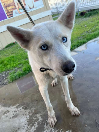
“Annie.” Photo courtesy of Clearlake Animal Control. This week’s dogs include “Annie,” a female Siberian husky mix with a white coat and blue eyes.
There also is “Rose,” a female pit bull terrier mix with a copper and white coat.
“Rose.” Photo courtesy of Clearlake Animal Control. The shelter is located at 6820 Old Highway 53. It’s open from 9 a.m. to 6 p.m. Tuesday through Saturday.
For more information, call the shelter at 707-762-6227, email This email address is being protected from spambots. You need JavaScript enabled to view it., visit Clearlake Animal Control on Facebook or on the city’s website.
This week’s adoptable dogs are featured below.
Email Elizabeth Larson at This email address is being protected from spambots. You need JavaScript enabled to view it.. Follow her on Twitter, @ERLarson, or Lake County News, @LakeCoNews.
UC San Francisco researchers have designed a candidate drug that could help make pancreatic cancer, which is almost always fatal, a treatable, perhaps even curable, condition.
The new drug candidate permanently modifies a wily cancer-causing mutation, called K-Ras G12D, that is responsible for nearly half of all pancreatic cancer cases and appears in some forms of lung, breast and colon cancer.
Pancreatic cancer is less common than these other cancers, but the lack of treatment options makes it more deadly, and it claims more than 50,000 lives each year in the United States.
“We’ve worked for 10 years to bring pancreatic cancer therapies up to speed with therapies for other cancers,” said Kevan Shokat, PhD, a professor in the Department of Cellular and Molecular Pharmacology who led the work. “This breakthrough is the first to target G12D and gives us a firm foothold to fight this devastating mutation.”
The findings appear March 5, 2024, in Nature Chemical Biology.
Shokat and his colleagues developed the first cancer drugs to stop a different K-Ras mutation, G12C, in 2013. Since then, two therapies have been approved for use in lung and breast cancer, but the advance didn’t move the needle for treating pancreatic cancer.
An extremely common mutation
K-Ras mutations are extremely common in pancreatic cancer, explaining 90% of cases. About half of these mutations are G12D, which differs from most other K-Ras mutations by a single amino acid substitution.
This difference between healthy and cancer-causing proteins, in which glycine (G) becomes aspartate (D), presented a monumental challenge for chemists.
“There are very few molecules out there that can sense the difference between the cancer-causing aspartate and the glycine,” Shokat said. “To make good therapies, we need drugs that work on the tumor cells only, without affecting healthy cells.”
Shokat’s team envisioned a molecule that fit into a pocket of the K-Ras protein, then firmly – and irreversibly – bound to the rogue aspartate. The explosion of research that followed Shokat’s 2013 discovery enabled them to develop a template for chemicals that reliably found their way into that corner of the protein.
“Once we had that structure for our molecules, we knew they were sitting in the protein at the right spot,” Shokat said. “Then we could explore the little nooks and crannies that we needed to discover the chemistry of the aspartate.”
Could a bend in a molecule lead to a cure?
The scientists went through dozens of chemicals.
“It’s like climbing a new route on a mountain, you may be strong but the lengths of your arms limit what you can do,” Shokat said. “It was a lot of trial and error, tweaking the branches of these molecules to position them in this incredibly tight space around G12D. Some got close, then failed, and we would start over.”
Eventually, they found a winning molecule. It settled into the appropriate corner of K-Ras and bent into a new shape that reacted strongly with the aspartate.
The molecule put the brakes on tumor growth from G12D in cancer cell lines, as well as an animal model of human cancer. And it never attacked healthy proteins.
The scientists are now optimizing the molecule to be durable enough to fight cancer in the human body. With the traction gained from this study, Shokat said, new therapies for pancreatic cancer could enter clinical trials in as little as two to three years.
“We’ve learned a lot from other targeted therapies and know how to quickly translate discoveries like these for the clinic,” said Margaret Tempero, MD, director of the UCSF Pancreas Center. “An effective drug targeting K-RAS G12D could be transformative for patients with pancreatic cancer.”
For funding and disclosures, see the paper. Other UCSF authors are Quinheng Zheng, Ziyang Zhang, and Keelan Z. Guiley. Zhang is now a professor at UC Berkeley.
Levi Gadye writes for the University of California, San Francisco.
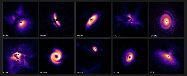
The images shown here were captured using the Spectro-Polarimetric High-contrast Exoplanet REsearch (SPHERE) instrument mounted on ESO’s Very Large Telescope (VLT). SPHERE’s state-of-the-art extreme adaptive optics system corrects for the turbulent effects of Earth’s atmosphere, yielding crisp images of the discs around stars. The stars themselves have been covered with a coronagraph — a circular mask that blocks their intense glare, revealing the faint discs around them. The discs have been scaled to appear roughly the same size in this composition. Credit: ESO/C. Ginski, A. Garufi, P.-G. Valegård et al.
In a series of studies, a team of astronomers has shed new light on the fascinating and complex process of planet formation. The stunning images, captured using the European Southern Observatory's Very Large Telescope (ESO’s VLT) in Chile, represent one of the largest ever surveys of planet-forming discs. The research brings together observations of more than 80 young stars that might have planets forming around them, providing astronomers with a wealth of data and unique insights into how planets arise in different regions of our galaxy.
“This is really a shift in our field of study,” says Christian Ginski, a lecturer at the University of Galway, Ireland, and lead author of one of three new papers published today in Astronomy & Astrophysics. “We’ve gone from the intense study of individual star systems to this huge overview of entire star-forming regions.”
To date more than 5000 planets have been discovered orbiting stars other than the Sun, often within systems markedly different from our own Solar System. To understand where and how this diversity arises, astronomers must observe the dust- and gas-rich discs that envelop young stars — the very cradles of planet formation. These are best found in huge gas clouds where the stars themselves are forming.
Much like mature planetary systems, the new images showcase the extraordinary diversity of planet-forming discs. “Some of these discs show huge spiral arms, presumably driven by the intricate ballet of orbiting planets,” says Ginski. “Others show rings and large cavities carved out by forming planets, while yet others seem smooth and almost dormant among all this bustle of activity,” adds Antonio Garufi, an astronomer at the Arcetri Astrophysical Observatory, Italian National Institute for Astrophysics (INAF), and lead author of one of the papers.
The team studied a total of 86 stars across three different star-forming regions of our galaxy: Taurus and Chamaeleon I, both around 600 light-years from Earth, and Orion, a gas-rich cloud about 1600 light-years from us that is known to be the birthplace of several stars more massive than the Sun. The observations were gathered by a large international team, comprising scientists from more than 10 countries.
The team was able to glean several key insights from the dataset. For example, in Orion they found that stars in groups of two or more were less likely to have large planet-forming discs. This is a significant result given that, unlike our Sun, most stars in our galaxy have companions.
As well as this, the uneven appearance of the discs in this region suggests the possibility of massive planets embedded within them, which could be causing the discs to warp and become misaligned.
While planet-forming discs can extend for distances hundreds of times greater than the distance between Earth and the Sun, their location several hundreds of light-years from us makes them appear as tiny pinpricks in the night sky.
To observe the discs, the team employed the sophisticated Spectro-Polarimetric High-contrast Exoplanet REsearch instrument, or SPHERE, mounted on ESO’s VLT. SPHERE’s state-of-the-art extreme adaptive optics system corrects for the turbulent effects of Earth’s atmosphere, yielding crisp images of the discs.
This meant the team were able to image discs around stars with masses as low as half the mass of the Sun, which are typically too faint for most other instruments available today.
Additional data for the survey were obtained using the VLT’s X-shooter instrument, which allowed astronomers to determine how young and how massive the stars are. The Atacama Large Millimeter/submillimeter Array, or ALMA, in which ESO is a partner, on the other hand, helped the team understand more about the amount of dust surrounding some of the stars.
As technology advances, the team hopes to delve even deeper into the heart of planet-forming systems. The large 39-meter mirror of ESO’s forthcoming Extremely Large Telescope (ELT), for example, will enable the team to study the innermost regions around young stars, where rocky planets like our own might be forming.
For now, these spectacular images provide researchers with a treasure trove of data to help unpick the mysteries of planet formation.
“It is almost poetic that the processes that mark the start of the journey towards forming planets and ultimately life in our own Solar System should be so beautiful,” concludes Per-Gunnar Valegård, a doctoral student at the University of Amsterdam, the Netherlands, who led the Orion study.
Valegård, who is also a part-time teacher at the International School Hilversum in the Netherlands, hopes the images will inspire his pupils to become scientists in the future.
LAKE COUNTY, Calif. — The Lake County Registrar of Voters Office on Thursday offered an update on the work to complete the official canvass for the March 5 presidential primary.
The 28-day canvass period began the day after the election. Until it is complete and the election is certified, the election results are not final.
Since the last update, the March 12 deadline passed to receive ballots postmarked by the end of Election Day.
The elections office said that approximately 7,415 vote-by-mail, or absentee, ballots remain to be counted. That’s about 600 less than the last count, which was impacted by those additional ballots continuing to come in by mail.
There also are 266 provisional or conditional ballots, and 266 vote-by-mail ballots that require further review for various reasons, the agency reported.
The elections office said the grand total of ballots remaining to be counted as of Thursday was 7,947.
Effective this year, AB 63 requires that the elections office update vote results and unprocessed ballot counts at least once per week and post the updated information on its website.
Registrar of Voters Maria Valadez said her office may stop posting results if the only ballots left to count are the ballots for which voters have the opportunity to either verify their signature or provide a signature, or until they certify election results.
For more information, visit the Lake County Registrar of Voters website or call 707-263-2372 OR toll-free at 888-235-6730.
Editor’s note: This story has been corrected to state that the canvass period is 28 days, not 30, and to add information about AB 63.
 How to resolve AdBlock issue?